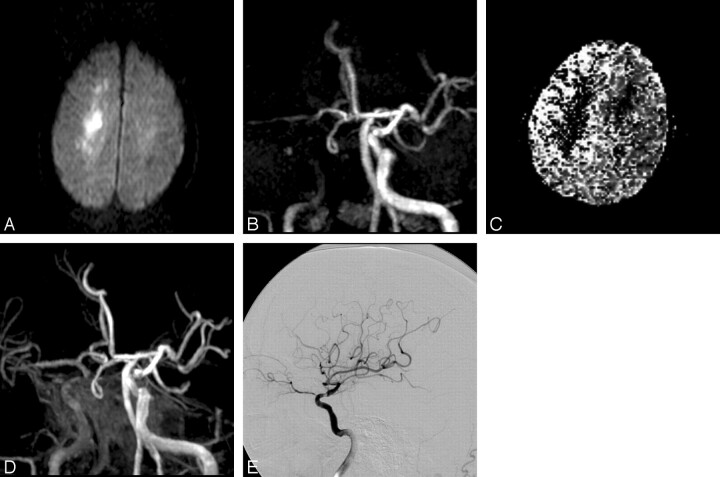
Fig 1.
A 51-year-old man with progressive left-sided weakness, dysarthria, and impaired consciousness (patient 4). A, Axial DWI confirms infarction in the centrum semiovale. B, On 3D TOF MR angiogram (anteroposterior [AP] view), occlusion of the C4 segment of the right ICA is suspected. Antegrade filling of the MCA via an anterior communicating artery is identified. C, Relative mean transit time map shows a region of delayed flow in the right hemisphere. D, Depiction of the cavernous portion of the ICA is improved on postcontrast 3D TOF MR angiogram (AP view), which shows severe stenosis at the C2 portion of the ICA. Some contrast enhancement from the cavernous sinus is evident. E, Right internal carotid angiogram immediately after MR imaging examination shows severe stenosis at the C2 portion and delayed flow in the MCA territory, but no antegrade filling into the anterior cerebral artery territory (TIMI grade 1).

