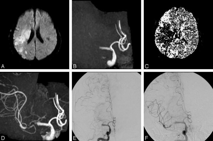
Fig 2.
A 49-year-old man with sudden-onset left-sided paralysis, aphasia, and impaired consciousness (patient 6). A, Axial DWI obtained at the time of MRA confirms infarction of right MCA territory. B, 3D TOF MR angiogram (AP view) shows occlusion of the M1 segment of the right MCA. C, Relative mean transit time map shows a region of delayed flow in the right hemisphere. D, Postcontrast 3D TOF MR angiogram (AP view) shows improved depiction of the distal M1 portion and M2 branches. Some contrast enhancement from the cavernous sinus is evident. E, Right internal carotid angiogram immediately after MR imaging examination shows total occlusion (TIMI grade 0) of the M1 segment of the right MCA. Retrograde opacification of the MCA branches via pial collateral vessels extending from the anterior cerebral artery is noted; however, the vessel just distal to the occlusion is not delineated. F, Right internal carotid angiogram after the first trial of angioplasty by using the 9-mm-long balloon catheter at the occluded portion shows antegrade filling into the distal M1 portion and M2 branches through the residual stenosis. Note that postcontrast MR angiogram (D) precisely demonstrates the extent of the occlusion that is shown on catheter angiography during PTCBA procedures (F).

