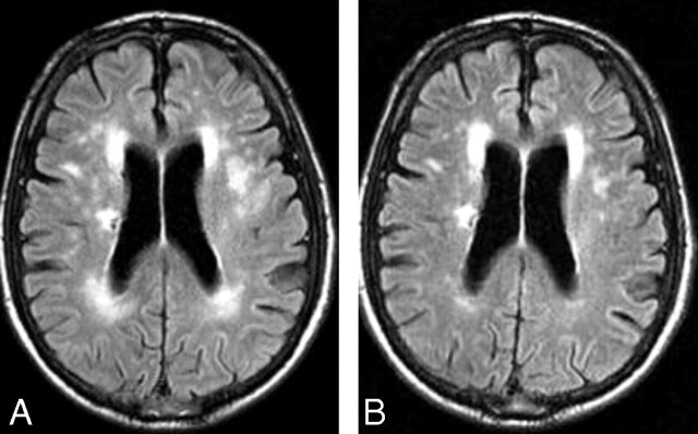
Fig 3.
A, MR image (transverse T2-weighted fast-FLAIR images, 9900 ms/110 ms/2500 ms/1 [TR/TE/inversion time/acquisitions]) obtained in patient 3 at baseline, while showing signs of overt hepatic encephalopathy. MR shows multiple focal WMLs (total lesion volume, 18,601 mm3). B, Patient 3 after improvement of hepatic encephalopathy: MR shows clear reduction in size and number of focal WMLs (final lesion volume, 10,393 mm3).

