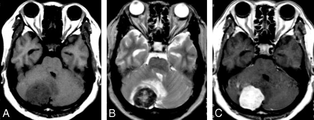
Fig 2.
A 52-year-old man with headache and dizziness for 6 months. A, T1-weighted axial MR image, a homogeneous low signal intensity mass is noted in the right cerebellum. B, T2-weighted axial MR image shows 2 different signal intensity portions of the mass: the peripheral hyposignal intensity and the central hypersignal intensity to gray matter. Mass effect on the fourth ventricle is also noted due to peritumoral edema. C, Gadolinium-enhanced T1-weighted axial MR image shows marked homogenous enhancement.

