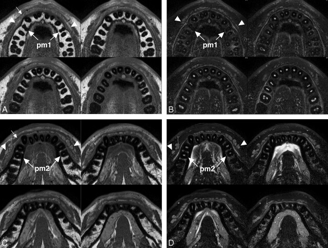
Fig 1.
A 26-year-old man with healthy upper and lower labial glands. A, Sequential (upper left to lower right panels in a cephalocaudal direction) axial T1-weighted images using a 47-mm microscopy coil (TR/TE/number of signal-intensity acquisitions, 550/10 msec/3; section thickness, 2 mm; section gap, 0.2 mm) of the upper labial salivary gland (arrowheads) show gland clusters spanning the left and right first premolar (pm1) regions. Small arrow indicates orbicularis oris muscle. B, Sequential (upper left to lower right panels in a cephalocaudal direction) axial fat-suppressed T2-weighted images using a microscopy coil (TR/TE/number of signal-intensity acquisitions, 3140/90 msec/4) of the upper labial salivary gland (arrowheads) show hyperintense gland clusters consisting of 1 layer in the anterior part and, at most, 2 layers in the posterior part of the lip. pm1 indicates the first premolar. C, Sequential (upper left to lower right panels in a cephalocaudal direction) axial T1-weighted images using a microscopy coil (TR/TE/number of signal-intensity acquisitions, 550/10 msec/3) of the lower labial gland (arrowheads) show gland clusters spanning the left and right second premolars (pm2). Small arrow indicates the orbicularis oris muscle. D, Sequential (upper left to lower right panels in a cephalocaudal direction) axial fat-suppressed T2-weighted images using a microscopy coil (TR/TE/number of signal-intensity acquisitions, 3140/90 msec/4) of the lower labial salivary gland (arrowheads) show hyperintense gland clusters consisting of, at most, 2 layers in the anterior part and of, at most, 2 or 3 layers in the posterior part of the lip. pm2 indicates the second premolar.

