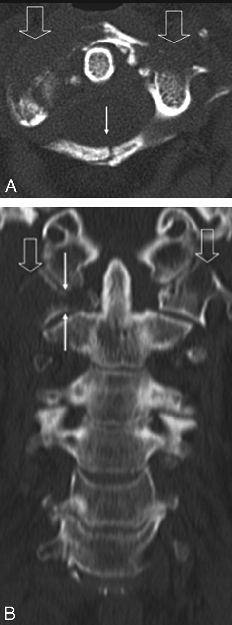
Fig 3.

A, A 50-year-old woman with history of lung carcinoma with heavy neck pain after minor blunt cervical trauma. Axial 2.5-mm image shows bilateral osteolytic lesions in each lateral mass of the atlas (C1) (open arrows) and midline fracture (arrow) of the posterior arc of C1. Calculated effective dose of low-dose 6-MDCT examination (130 kV and 89 mAs) is 1.48 mSv. B, Coronal 2.5-mm image confirms bilateral osteolytic lesions in each lateral mass of C1 (open arrows) and transverse fracture (arrows) trough the right lateral mass.
