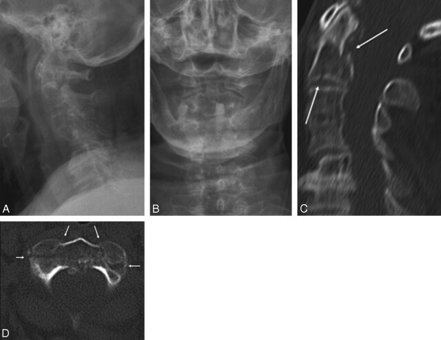
Fig 6.
A, An 82-year-old woman with acute neck pain after a motor vehicle crash. Standard radiography, lateral view, was interpreted as negative, but additional CT was proposed because of technical failure to view the lower cervical segment C7 (superposition of the shoulders). B, Standard radiograph with odontoid view was interpreted as negative for fracture in the region of the cranio-cervical junction. C, Sagittal 2.5-mm image of low-dose 16-MDCT-examination (100 kV and 165 mAs) clearly depicts fracture (arrows) at the base of the axis (C2). D, Axial 2.5-mm image of the same low-dose CT examination shows more complex fracture of the body of C2, bilaterally extending in the lateral masses (arrows). Calculated effective dose of MDCT examination is 1.3 mSv.

