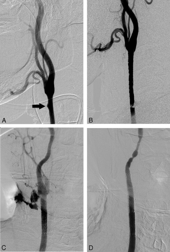
Fig 2.

A 69-year-old man with a small, blisterlike lesion of the right common carotid artery, detected incidentally during surgical reconstruction of his wound. A, Conventional angiography shows a small ulcerative lesion (arrow) in the distal portion of the right common carotid artery. B, The lesion disappeared after preventive placement of the covered stent. C, A massive contrast extravasation is noted, near the distal end of the previous inserted stent, on the right common carotid angiogram obtained for the evaluation of the rebleeding, which occurred 3 weeks after the initial procedure. D, Complete control of hemorrhage is achieved by additional insertion of a covered stent.
