
Fig 3.
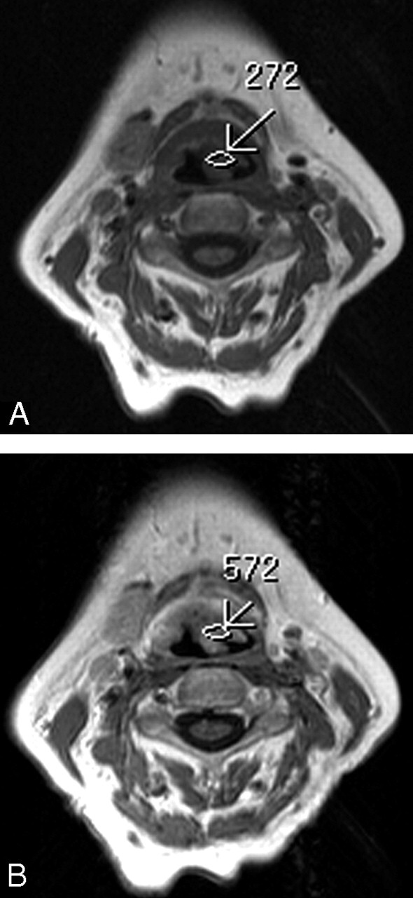
Axial T1-weighted MR images (600/15 ms [TR/TE]) before (A) and after (B) contrast administration show a supraglottic mass with the SI that increases from 246 to 513 within the tumor area (arrows). The degree of contrast enhancement was (572–272)/272 = 110%. The tumor was considered a high-enhanced tumor. Local recurrence was not documented within 24 months after RT.