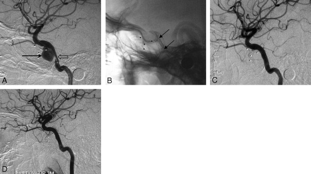
Fig 3.
Case 6, a 23-year-old man with a pseudoaneurysm secondary to post-balloon embolization of a CCF. A, Lateral cerebral angiogram reveals a wide-necked pseudoaneurysm (black arrow) on the left C4 segment, with stenosis at the proximal part of the parent artery (empty arrow). B, Plain film after stent placement clearly shows the covered stent bridging the pseudoaneurysm and the stenosis (arrows). C, Lateral cerebral angiogram shows complete resolution of the aneurysm immediately after the stent placement. D, Cerebral angiography 3 months after the procedure shows total obliteration of the aneurysm with patency of the parent artery.

