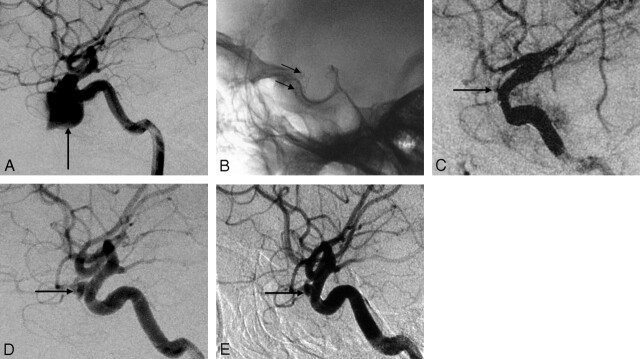
Fig 4.
Case 5, a 35-year-old man with massive epistaxis. A, Lateral cerebral angiogram shows a giant pseudoaneurysm on the left C5 segment (arrow). B, The Willis covered stent can be clearly seen in the plain film (arrows) after stent placement. C, Cerebral angiogram immediately after stent placement demonstrates a minimal endoleak into the pseudoaneurysm (arrow) in the orifice of the ophthalmic artery (arrow). D, Follow-up cerebral angiogram 2 months after the procedure demonstrates that retention of contrast medium at the orifice of the ophthalmic artery is increased (arrow), which suggests the existence of a residual cavity. E, Follow-up cerebral angiogram 6 months after the procedure demonstrates obvious shrinkage of the residual cavity (arrow) with patency of the parent artery.

