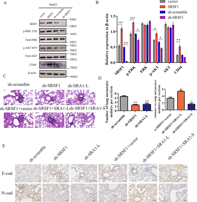
Fig. 6. In vivo, SRSF1 promotes the metastasis of HCC cells.
A, B The effect of SRA1 on downstream CD44, AKT, and ERK signaling pathway proteins was detected in HepG2 cells by western blot. C, D HE staining was used to analyze the number of pulmonary metastases in different groups of nude mice. The magnification of HE is 200× and the scale length is 50 μm. E The expression level of N-cad and E-cad in tumor tissues was analyzed by immunohistochemistry. The magnification of immunohistochemistry is 100× and the scale length is 100 μm. Data are presented as mean ± S.D. (N = 3). The “*, **, ***” indicate “P < 0.05, 0.01, 0.001” versus the control group, respectively.

