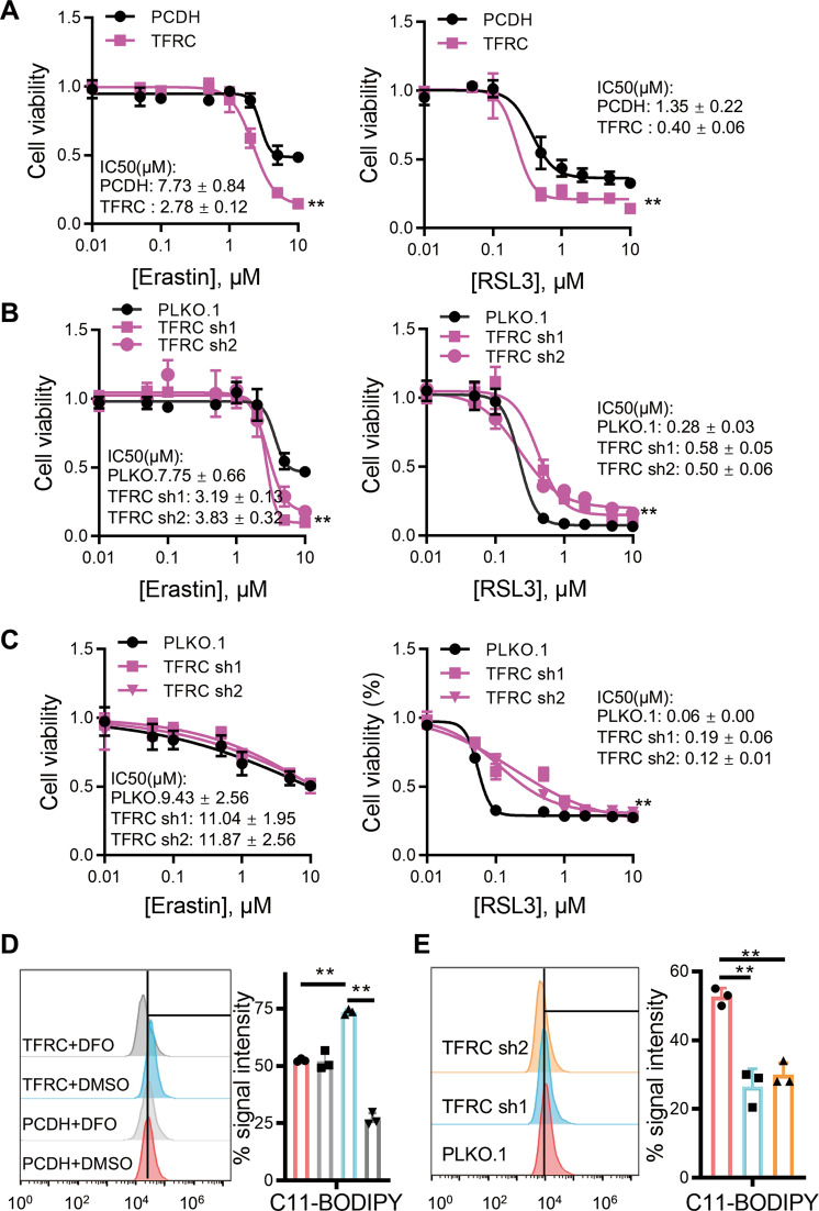
Fig. 5. TFRC mediates MYCN–induced, GPX4-dependent ferroptosis.
A Cell viability of SHEP cells with or without TFRC overexpression incubated with Erastin (left) or RSL3 (right). Cell viabilities of SHEP (B) or SK-N-BE2 (C) cells with or without TFRC depletion incubated with Erastin (left) or RSL3 (right). Amounts of cellular lipid peroxides measured by C11-BODIPY in SHEP (D) or SK-N-BE2 (E) cells with or without TFRC. Data are presented as mean ± SD of three replicates. Two-tailed unpaired t-tests were performed to calculate p values. **p < 0.01.

