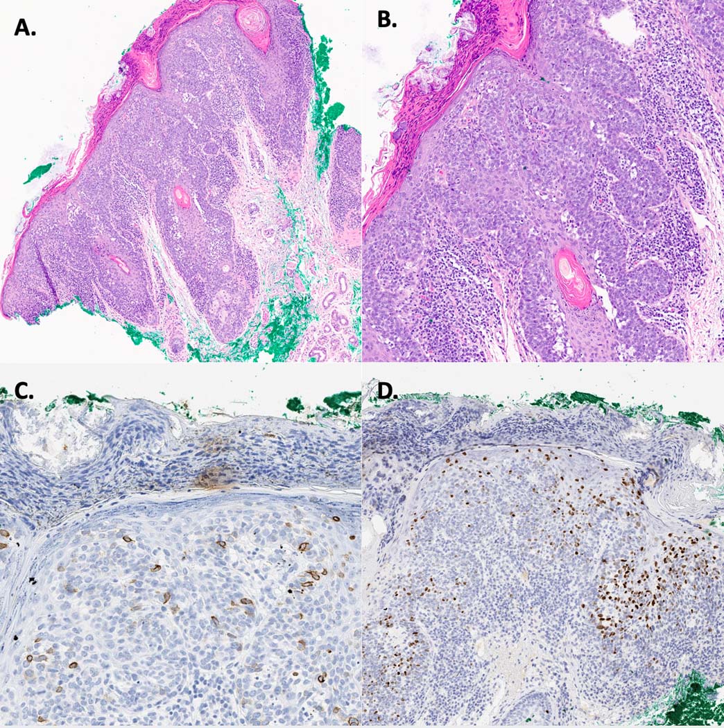
Figure 2:

Intraepidermal Merkel cell carcinoma. A. Photomicrograph showing an intraepidermal proliferation of cells with widespread pagetosis (H&E, 10X). B. Photomicrograph showing an intraepidermal proliferation of cells with widespread pagetosis; the cells are made of hyperchromatic epitheloid cells with a high mitotic rate, small nucleoli, and scant cytoplasm, all neuroendocrine features (H&E, 20X). C. Cells were positive for CK20. D. Cells were positive for INSM1.
