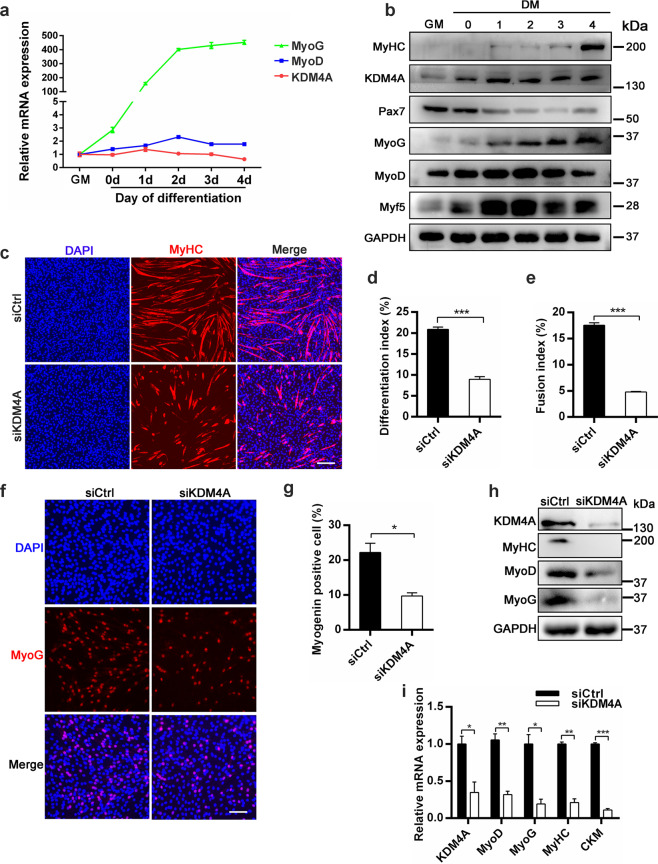
Fig. 4. KDM4A deficiency impedes myoblasts differentiation.
a mRNA expression profiles of KDM4A, MyoD and MyoG in C2C12 cells during differentiation. C2C12 cells were cultured in either growth medium for 2 days (GM, proliferating) or differentiation medium for 0, 1, 2, 3 or 4 d (DM 0-4). b Western blot analysis of C2C12 cells from GM to DM 4d was performed for the expression of KDM4A or myogenic genes and GAPDH served as loading control. c Immunofluorescence staining of MyHC for C2C12 cells in differentiation medium for 3 days. C2C12 cells were transfected with negative control siRNA (siCtrl) or KDM4A siRNA (siKDM4A) for 12 h in growth medium and then cultured in differentiation medium for 3 days. Scale bar = 200 μm. d, e Quantification of differentiation index and fusion index shown in the (c) (n = 3). f Representative images of immunofluorescence staining for MyoG in differentiated C2C12 cells. Cell nuclei were stained with DAPI. C2C12 cells were transfected with siCtrl or siKDM4A for 12 h and then induced to differentiate for 1 day that were stained for MyoG. Scale bar = 100 μm. g The number of MyoG+ cells was quantified showed that KDM4A knockdown inhibited myoblast differentiation. h Western blot analysis of MyoD, MyoG and MyHC protein levels in siCtrl and siKDM4A C2C12 cells after 3 d in differentiation medium. i mRNA expression of myogenic genes in siCtrl and siKDM4A C2C12 cells for 3 days differentiation (n = 3). Data are represented as mean ± SD. *P < 0.05; **P < 0.01; ***P < 0.001 (Student’s t test).

