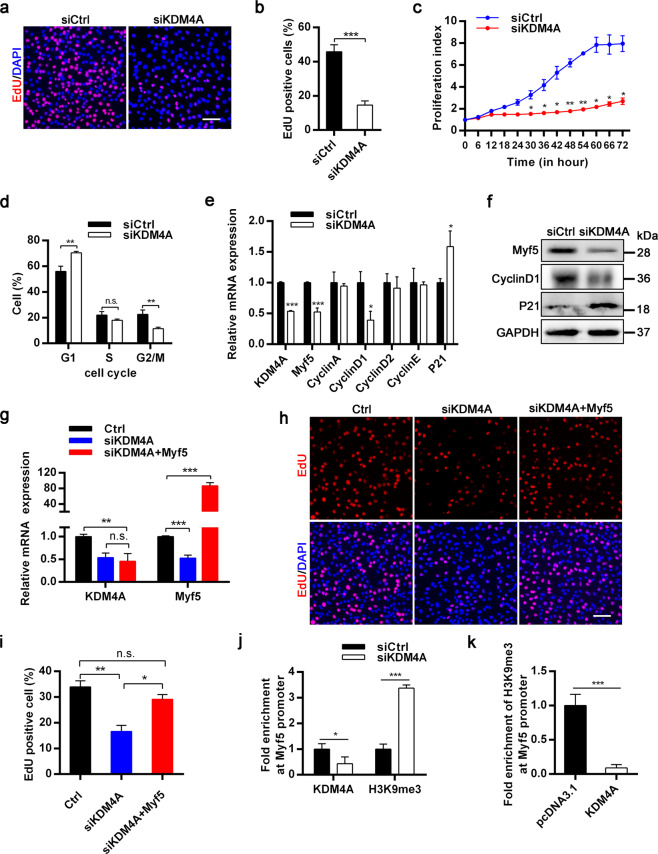
Fig. 7. KDM4A facilitates myoblasts proliferation by demethylating H3K9me3 at Myf5 loci.
a Representative images of the EdU staining for siCtrl or siKDM4A C2C12 cells on 36 h post-transfection. Scale bar = 100 μm. b Quantification of the percentage of EdU-positive cells in (a). c Real-time cell proliferation monitoring assay of the proliferation index of C2C12 cells. d The cell cycle distribution of proliferating myoblasts was analyzed through flow cytometry after PI staining. C2C12 cells were transfected with siCtrl and siKDM4A for 36 h. e Expression analysis of cell-cycle related genes in siCtrl and siKDM4A C2C12 cells in growth medium using qRT-PCR. f Western blot showing Myf5, Cyclin D1, P21 and GAPDH levels in the same conditions as in (e). g qRT-PCR showing the mRNA expression levels of KDM4A and Myf5 in proliferating C2C12 cells transfected with siRNA or plasmids as indicated. h EdU staining of C2C12 cells cotransfected with siCtrl or siKDM4A and control or Myf5 plasmid for 36 h in growth medium. i The number of EdU-positive cells was counted. j ChIP-qPCR analysis of the binding of KDM4A and H3K9me3 at My5 promoter in siCtrl and siKDM4A C2C12 cells. k The enrichment of H3K9me3 at Myf5 locus in C2C12 myoblasts transduced with either empty plasmid or KDM4A plasmid. Data are represented as mean ± SD. *P < 0.05; **P < 0.01; ***P < 0.001; n.s. not significant (Student’s t test).

