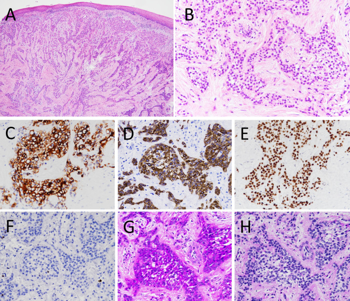
Fig. 2.
Histological and immunohistochemical findings in the biopsy specimen. a The tumor did not involve the overlying oral epithelium and showed typical histologic appearance of clear cell carcinoma (CCC). b High power view showed proliferation of epithelial cells with clear to eosinophilic cytoplasm. c AE1/AE3, d CK5/6 and e p63 were immunopositive. f MIB1 index was approximately 2%. g PAS was positive and h PAS diastase was negative

