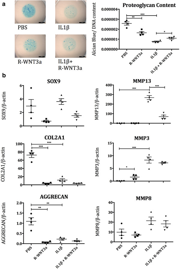
FIGURE 1.

a) HAC micromasses were treated with combinations of recombinant WNT3a (50 ng/ml R‐WNT3a) and IL1‐β (10 ng/ml) over 6 days, then stained with alcian blue (Ab) dye to assess proteoglycan content, which was normalised to DNA content to account for proliferation (n = 4). b) HAC were treated with IL1‐β (10 ng/ml) and R‐WNT3a (100 ng/ml) and mRNA readouts of cartilage anabolism and IL1‐β pathway activation assessed by QPCR after 24 h (n = 4)
