
FIGURE 3.
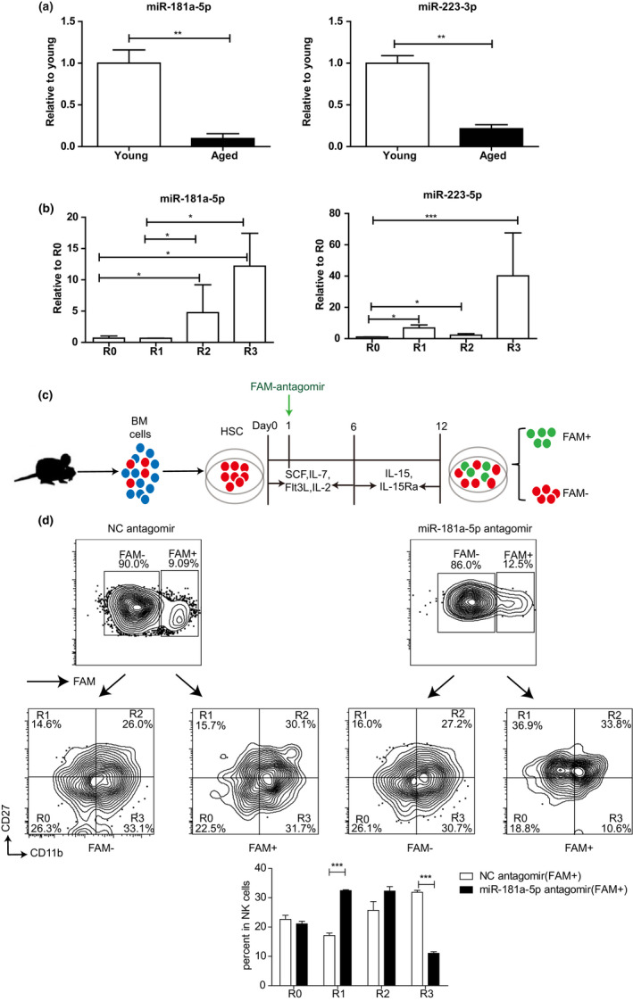
The declined expression of miR‐181a‐5p influences the maturation of NK cells in aged mice. (a) The expression levels of miR‐181a‐5p (left) and miR‐223‐3p (right) in CD 27+ NK cells of young or aged mice were detected using qRT‐PCR. The cells were sorted from bone marrow of the young and aged mice by using a FACSAria II (BD Biosciences) with purities >98%. (b) The expression levels of miR‐181a‐5p (left) and miR‐223‐3p (right) in different NK cell subsets of young mice. The cells were sorted from spleen, peripheral lymph nodes and bone marrow of the young mice. (c) Schematic of in vitro mouse NK cell differentiation using a two‐step cell culture system as described in Materials and Methods. HSCs purified from bone marrow of young CD45.1 mice were transfected with FAM‐labeled miR‐181a‐5p antagomir or FAM‐labeled NC antagomir. (d) Flow cytometry (above) and statistical results (below) of R0, R1, R2 and R3 NK cells in (c). Data were from three independent experiments (mean ± SEM). *p < 0.05, **p < 0.01, ***p < 0.001