
FIGURE 2.
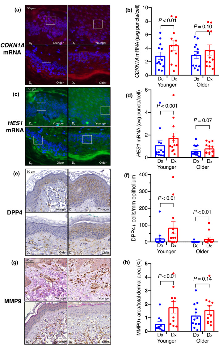
Representative images demonstrating CDKN1A mRNA (a), HES1 mRNA (c), and immunohistochemistry of senescence markers DPP4 (e) and MMP9 (g) in normal skin at baseline (D0) and after wounding (DX). CDKN1A mRNA signal (pseudo‐colored red); HES1 signal (pseudo‐colored green); DAPI (blue) as the nuclear stain. Each transcript is represented by a single punctum; the expression of each is represented as the average number of puncta per cell. Boxed areas are enlarged 2.5× in supplemental Figure S3. Both CDKN1A and HES1 mRNA were induced in response to wounding in the younger (p < 0.01, p < 0.001), but not the older (p = 0.10, p = 0.07) group (b,d). Images in (a) and (c) are 63X magnification. DPP4 levels (number of positive cells per millimeter of epithelium) were significantly induced in wounded skin in both the younger and older group (p < 0.01 for both) (f). MMP9 levels (proportion of positive area in the papillary dermis), in response to wounding, were significantly higher in the younger (p < 0.01) but not older (p = 0.14) group (h). Images in (e) and (g) are 40× magnification