
FIGURE 2.
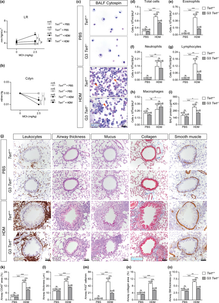
Telomerase deficiency attenuates AHR, eosinophil, and lymphocyte counts in BALF and reduces leukocyte presence and airway remodeling after HDM exposure. Quantification of lung resistance (LR) (a) and dynamic compliance (Cdyn) (b) to methacholine (MCh) evaluated by plethysmography in Tert +/+ and G3 Tert −/− mice. Representative BALF cytospin preparations (May‐Grünwald Giemsa (MGG) (orange arrowheads indicate eosinophils)) (c) and quantification of total (d) and differential BALF cell counts for eosinophils (e), neutrophils (f), lymphocytes (g), and macrophages (h), and total protein concentration in BALF (i) of Tert +/+ and G3 Tert −/− mice. Representative images of proximal airways showing CD45 (brown) and H&E (left), PAS (pink) (center) and Masson (blue), and SMA (brown) (right) stainings and immunostainings (j) and quantification of airway CD45+ area (leukocytes) (k), airway thickness (l), airway PAS+ cells (m), airway collagen area (n) and airway smooth muscle (SM) thickness (o) in lung sections from Tert +/+ and G3 Tert −/− mice. Quantifications in lung sections were performed in 4 different bronchi in a random way. Data are expressed as mean ±SEM. *p < 0.05; **p < 0.01; ***p < 0.001 (Dunn‐Sidak multiple comparison test). The number of mice is indicated in each case