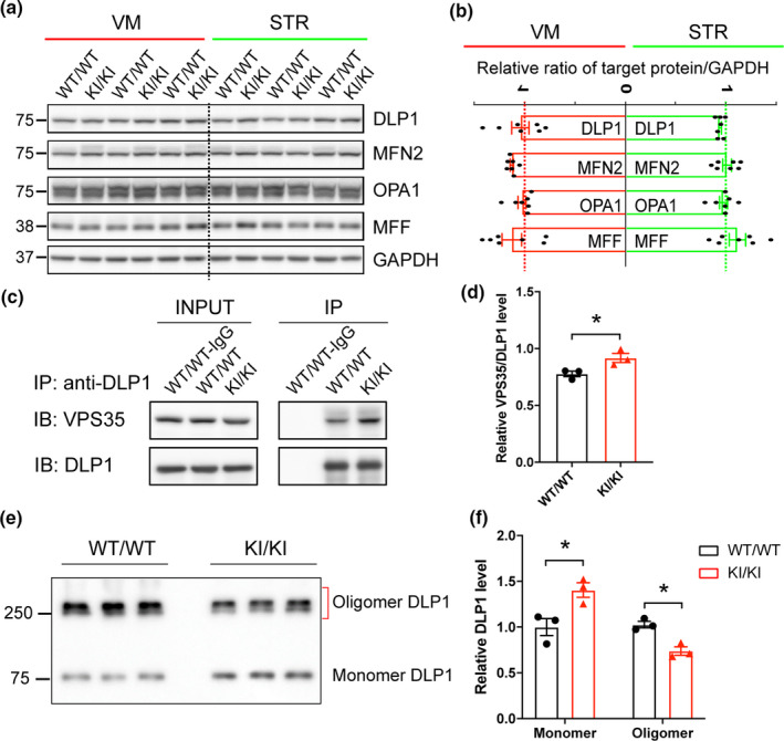
FIGURE 5.

Changes in mitochondrial fission/fusion proteins in VPS35 D620N KI mice. (a–b) Representative Western blots (a) and quantification (b) of mitochondrial fission/fusion proteins including DLP1, MFN2, OPA1, and MFF in VM and STR extracts from VPS35D620N / D620N and WT controls at 15‐month‐old (n = 7; Student's t test, unpaired, two‐tailed; data are shown as mean ± SEM; n.s., not significant). (c–d) Representative Western blot analysis (c) and quantification (d) of VPS35 in DLP1 immunoprecipitates from brain homogenates of VPS35D620N / D620N and WT mice (n = 3; Student's t test, unpaired, two‐tailed; data are shown as mean ± SEM; *p < 0.05). (e–f) Representative Western blot of DLP1 (e) and quantification (f) of oligomeric and monomeric DLP1 in the DSS‐treated mitochondrial fraction of VM from VPS35D620N / D620N and WT mice at 15‐month‐old (n = 3; unpaired t test; *p < 0.05)
