
FIGURE 3.
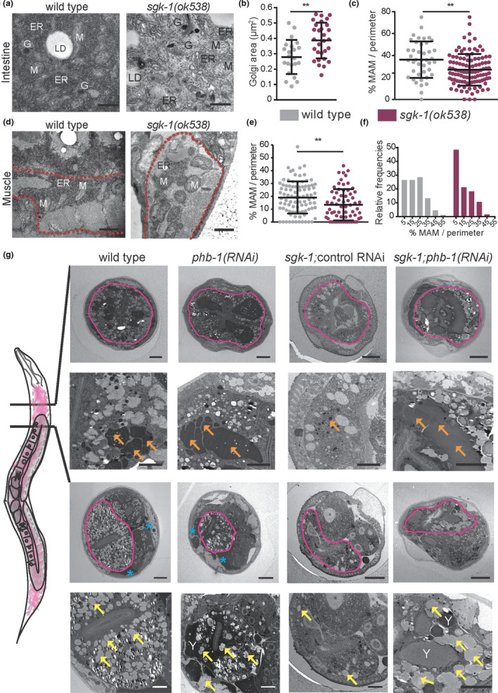
Prohibitin depletion suppresses lipogenesis and lipoprotein/yolk formation defects of sgk‐1 mutants. (a) Transmission electron microscopy (TEM) images of the first intestinal cells of wild‐type and sgk‐1 mutants at day one of adulthood. Bar size: 500 nm. ER: Endoplasmic Reticulum, LD: Lipid Droplet, Y: Yolk, G: Golgi. (b) Quantification of Golgi areas in intestine of wild‐type and sgk‐1 mutants at day one of adulthood. (c) Quantification of the ER‐mitochondria contact site length in intestine, normalized to mitochondrial perimeter, of wild‐type and sgk‐1 mutants, at day one of adulthood. Mean ± SD; **p < 0.05; t test, n > 40 contact sites. (d) TEM images of muscle, delineated with a red dashed line, of wild‐type and sgk‐1 mutants at day one of adulthood. Bar size: 1 μm. M: Mitochondria. (e) Quantification of the ER‐mitochondria contact site length in muscle, normalized to mitochondrial perimeter, of wild type and sgk‐1 mutants, at day one of adulthood. Mean ± SD; **p < 0.05; t‐test, n > 60 contact sites. (f) Frequency distribution histogram, as per cent, from data set shown in panel e, bin width 10. (g) TEM images of wild type, sgk‐1 mutants, phb‐1 depleted animals and sgk‐1;phb‐1(RNAi) treated mutants at day five of adulthood. Two sections, before and after the gonad turn are shown. Top images show a general view where the intestine is delineated with a pink line. Bottom images show a magnification of the intestinal area. Bar sizes: 10 μm (top panels) and 5 μm (bottom panels) for each of the cuts. Orange arrows mark yolk, blue asterisks mark pseudocoelomic lipoproteins, yellow arrows label LD, Y: yolk