Figure 1.
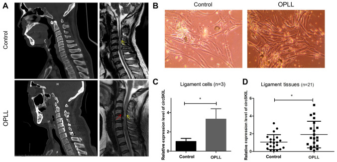
circSKIL is upregulated in OPLL cells and tissues. (A) Representative CT and MRI images of patients in the control and OPLL groups. Red arrow, OPLL lesion; yellow arrow, cervical spondylotic myelopathy lesion. (B) Morphology of primary cervical posterior longitudinal ligament cells. Magnification, x200. (C) circSKIL expression levels in OPLL and control isolated cells. (D) circSKIL expression levels in OPLL and control patient tissues. *P<0.05 vs. control. OPLL, ossification of the posterior longitudinal ligament; circ, circular RNA.

