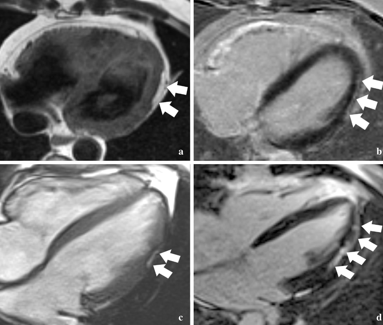
Fig. 1.

Patterns of left ventricular (LV) fat and fibrosis in arrhythmogenic right ventricular dysplasia/cardiomyopathy (ARVD/C). a Axial dark blood fast spin echo image obtained in a 46 year-old male proband with a PKP2 variant shows a typical bite-like pattern of epicardial fat infiltration in the apicolateral LV (arrows). b Axial late gadolinium enhancement (LGE) image in the same patient shows linear subepicardial enhancement suggesting fibrosis in the same location. c Axial balanced steady-state free precession cine image from a 28 year-old male proband with a PKP2 variant shows a typical “bite-like” pattern of LV fat infiltration on bright blood images, with the associated dark etching artifact between the fat and the myocardium. d Axial LGE image in the same patient shows more extensive enhancement indicating fibrosis in a predominantly linear subepicardial pattern along the lateral wall
