Figure 1.
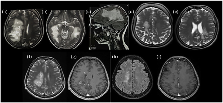
Radiological characteristics of two patients presented with Marburg-like tumefactive lesions. Bilateral Marburg-like tumefactive lesions of a patient at disease onset [(a)–(c)], after chronic treatment with mitoxantrone, cyclophosphamide, and glatiramer acetate showing significant resolution of the lesions [(d) and (e)]. This patient, 10 years after disease onset has an EDSS score of 1. A second patient with Marburg-like demyelination in the right centrum semiovale at onset [(f)] with Gd+ [(g)] and after three monthly cycles of cyclophosphamide. A significant resolution of the tumefactive lesion with no Gd+ was observed after three monthly cycles of cyclophosphamide [(h) and (i)] with EDSS score 1.5.
T2-weighted images: (a), (b), (d) to (f). FLAIR images: (c), (h). T1-weighted contrast-enhanced images: (g), (i).
EDSS, Expanded Disability Status Scale; FLAIR, fluid-attenuated inversion recovery; Gd+, gadolinium enhancement.

