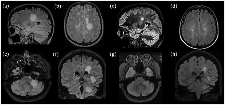
Figure 4.
Paradigms of successfully treated patients with TDL after alemtuzumab or rituximab. (a) and (b) Images represent a patient with a TDL in the left centrum semiovale, treated in the acute phase with IVMP. Subsequently, the patient developed a typical MS and received 6 months later the first course of alemtuzumab; an excellent clinical and radiological response 1 year after was noted with also a significant decrease in the TDL size [(c) and (d)]. The second patient presented with multiple TDLs in the left cerebral hemisphere and left middle cerebellar peduncle [(e) and (f)]; responded well to rituximab courses with no further clinical attacks [(g) and ( h)].
FLAIR images: (a), (b), (d) to (h). Double inversion recovery (DIR) image: (c).
Gd+, gadolinium enhancement; IVMP, intravenous methylprednisolone; MS, multiple sclerosis; TDL: tumefactive demyelinating lesion.

