Figure 5.
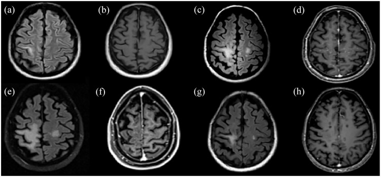
A paradigm of successfully treated TDL with rituximab in a patient with MS during chronic treatment with natalizumab. (a) and (b) The patient presented for the first time TDL-like lesion (non-PML) in the right precental gyrus with no Gd+ during natalizumab treatment. (c) and (d) After natalizumab withdrawal and IVMP course, a significant MRI activity with enlargement of the TDL (non-PML) and multiple bilateral hemispheric punctuate Gd+ lesions were noted. (e) and (f) Further increase in size of the TDL with a little reduction of the punctuate Gd+ lesions following three monthly cycles of cyclophosphamide (cyclophosphamide failure, 9 months after TDL-like lesion appearance). (g) and (h) Remarkable reduction of the TDL (non-PML) with slight remaining Gd+ post-rituximab initiation.
FLAIR images: (a), (c), (e), (g). T1-weighted contrast-enhanced images: (b), (d), (f), (h).
Gd+, gadolinium enhancement; IVMP, intravenous methylprednisolone; MRI, magnetic resonance imaging; MS, multiple sclerosis; PML, progressive multifocal leukoencephalopathy; TDL, tumefactive demyelinating lesion.

