Figure 3.
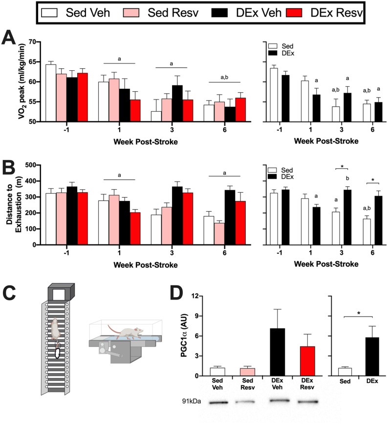
Stroke resulted in decreases in cardiovascular fitness, which was attenuated with delayed exercise. (A) Left panel: stroke reduced VO2 peak during a maximal exercise test (n = 5-9). Right panel: The exercise rehabilitation attenuated further reductions in VO2 peak (Time × DEx; n = 13-17). (B) Left panel: the distance rats could run until exhaustion was reduced at week 1 and week 6 poststroke (n = 5-9). Right panel: rats that engaged in the exercise rehabilitation could run farther until exhaustion at week 3 and week 6 poststroke compared with being sedentary (Time × DEx; n = 13-17). (C) The delayed exercise rehabilitation was a combination of resistance (ladder climbing) and aerobic (treadmill) exercise. (D) Left panel: PGC1α protein content in the red portion of the tibialis anterior (n = 7-8). Right panel: exercise increased PGC1α protein content, indicative of increased aerobic metabolism (n = 15-16). Lower panel: representative Western blot image.
Abbreviations: Resv, resveratrol; Sed Veh, sedentary vehicle control; Sed Resv, sedentary Resv; DEx Veh, exercise vehicle; DEx Resv, exercise Resv; VO2 peak, peak oxygen consumption; PGC1α, proliferator-activated receptor gamma coactivator 1-α.
a Different from prestroke.
bDifferent from 1 week poststroke.
*P < .05.

