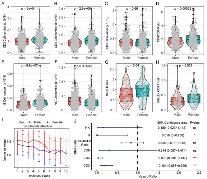
Figure 9.

The difference of lymphocytes in COVID-19 patients of different genders and their prognostic effects. (A) The proportion of CD3 T cells in PBMC of COVID-19 patients of different genders. (B) The proportion of CD4 T cells in PBMCs of COVID-19 patients of different genders. (C) Proportion of CD8 T cells in PBMC of COVID-19 patients of different genders. (D) Differences in the ratio of CD4/CD8 in PBMC of COVID-19 patients of different genders. (E) Proportion of B cells in PBMC of COVID-19 patients of different genders. (F) The proportion of NK cells in PBMC of COVID-19 patients of different genders. (G) The proportion of Naive B cells in PBMCs of COVID-19 patients of different genders. (H) Proportion of Effector CD8 T cells in PBMC of COVID-19 patients of different genders. Kruskal–Wallis test was used for statistical significance. (I) The cumulative event rate shows that lymphocyte absolute value contributes to the prognosis of all COVID-19 patients. (J) The cumulative event rate shows the contribution of lymphocyte absolute value to the prognosis of male COVID-19 patients. (K) The cumulative event rate shows the contribution of lymphocyte absolute value to the prognosis of female COVID-19 patients. (L) The line chart shows the dynamic changes of lymphocyte absolute value of COVID-19 patients of different genders. With the progress of treatment and the passage of time, multiple test values during the patient’s hospitalization are used for comparison.
