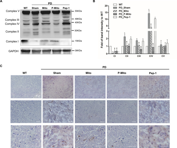
Figure 4.
Normalized expression profiles of mitochondrial complexes in lesioned substantial nigra (SN) document subsequent attenuation of oxidative stress. (A) Mitochondrial integrity in the SN of the midbrain after 3 months of treatment was assessed and (B) quantified based upon protein levels of mitochondrial respiratory chain complexes I–V in same PAGE gel, normalized against an internal control, glyceraldehyde 3-phosphate dehydrogenase (GAPDH), with same amount of loading protein. Changes in the patterns of mitochondrial proteins, including band intensity and molecular weights, relative to wild-type controls, are presented. For whole uncropped images of original western blots with three independent samples of each group, please refer to Supplementary Figure (Fig. S1). (C) Immunohistological stain for 8-hydroxy-2'-deoxyguanosine (8-OHdG), a marker of DNA oxidative damage, was performed to examine DNA oxidative damage in the SN. Brown color indicates specific immunostaining of 8-OHdG (as depicted by arrows at high magnification, lower panels). Blue dots indicate nuclear haematoxylin staining. +P < 0.05, vs. WT; *P < 0.05, vs. Sham; WT wild-type controls, PD Parkinson’s disease, Sham vehicle alone, Mito mitochondrial alone, P-Mito Pep-1-labelled mitochondria.

