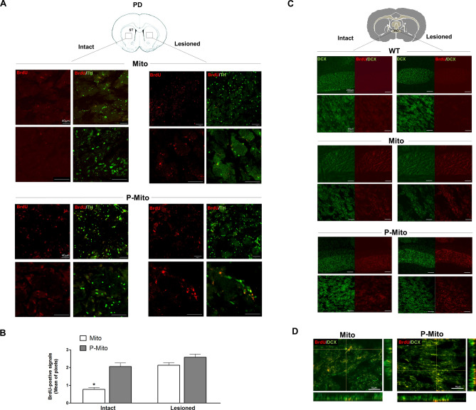
Figure 7.
BrdU-labeled mitochondria were expressed in striatal DA neurons and DCX-positive neurons of the anterior commissure (AC), defined by cross-sectional areas of the forebrain. (A) Double immunofluorescent staining of BrdU (red) with TH (green) was performed to confirm intranasally administered mitochondria in striatal DA neurons and (B) BrdU signals were quantified individually in lesioned and contralateral (intact) sides using ImageJ Software. (C) Reconfirmation of contralateral delivery of mitochondria employed double immunofluorescent staining, using BrdU (red) with DCX antibody (green) in the intact and lesioned sides of the AC, a bundle of axons connecting the two cerebral hemispheres. Images were taken at different magnifications in each group. (D) The merged image of Z-Stack confocal images, shown as side views of the x–z and y–z planes, revealed the localization of exogenous mitochondria in DCX-positive neurons. *P < 0.05, vs. Lesioned side, WT wild-type controls, Mito mitochondrial alone, P-Mito Pep-1-labelled mitochondria.

