Fig. 2.
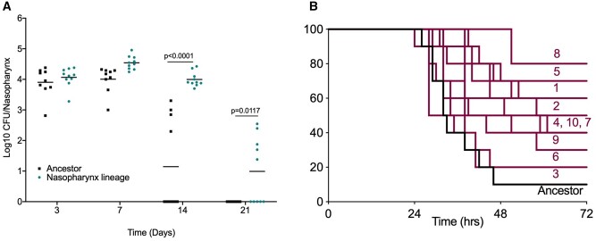
Tissue-passaged pneumococci show evidence of niche adaptation. Mice were infected with the ancestral D39 population or 20 times passaged lineages from (A) nasopharyngeal carriage (lineage 1) or (B) pneumonia models. (A) Mice were infected with 1 × 10⁵ CFU of the ancestor or nasopharynx lineage 1 to induce asymptomatic carriage. Bacterial numbers in nasopharynx were determined by serial dilution of tissue homogenates onto blood agar at days 3, 7, 14, and 21 postinfection. P-values are from two-way ANOVA with Sidak’s multiple comparison test. (B) Mice were infected with 1 × 10⁶ CFU of the ancestor or one of the ten lung-passaged lineages to induce pneumonia (n = 10 per group). Mice were monitored for signs of disease and culled once lethargic. Survival curves were compared by Mantel–Cox log-rank test (table 1). Labels show lineage numbers.

