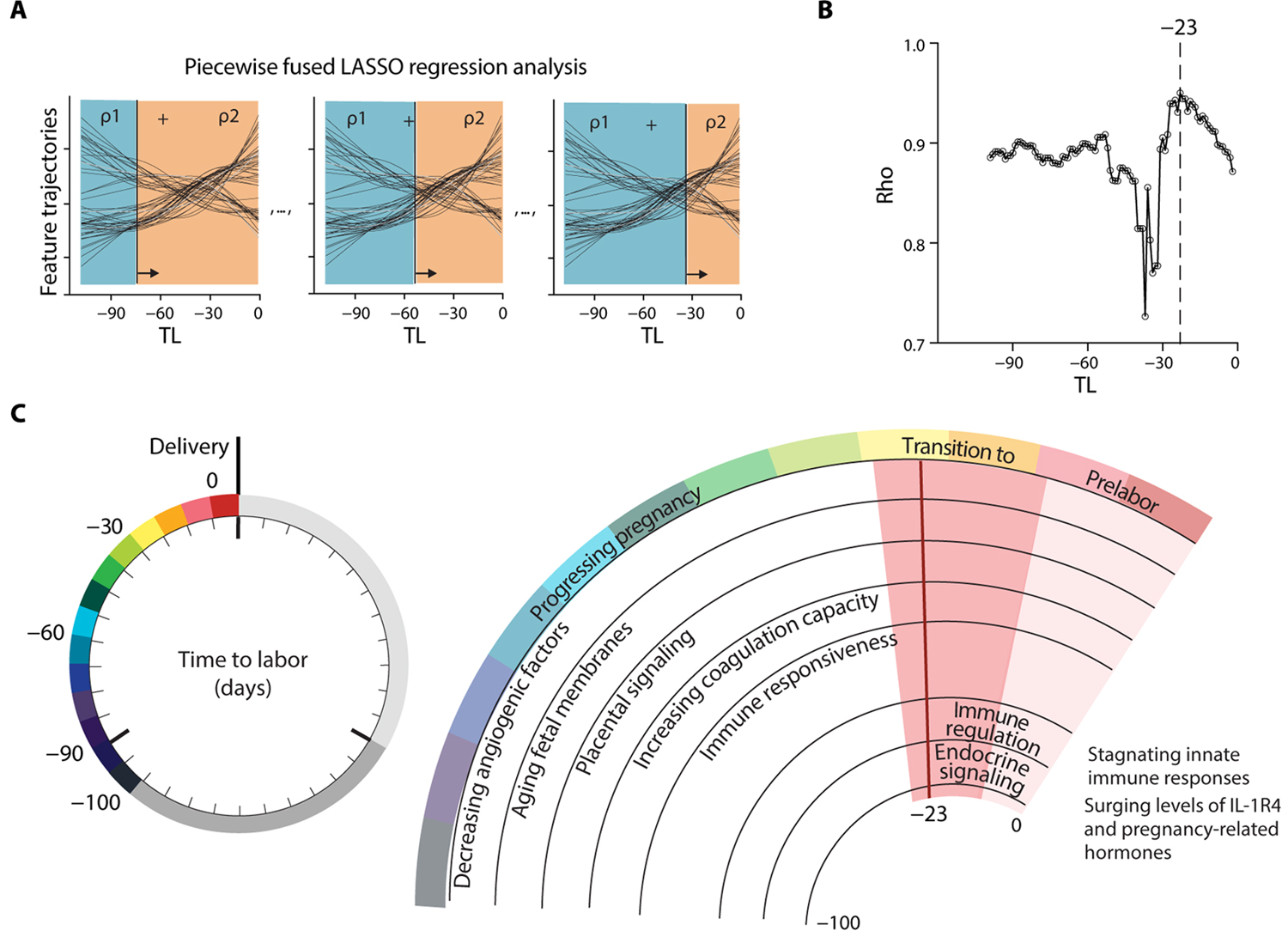
Fig. 5. A breakpoint in omic trajectories demarcates the transition from pregnancy maintenance to prelabor biological adaptations.

(A) Schematic of a piecewise fused LASSO regression combining predictions rho (ρ) of two regression models built from all datasets before and after a particular TL threshold, while sliding the threshold across the time axis. Plotting ρ over time reveals the time point of highest accuracy (maximum ρ). (B) Maximum ρ of 0.95 was observed at day −23 (range [−27, −13]; N = 53 patients). (C) Summary of concerted biological adaptations depicting a clock to labor. Angiogenic factors: Decreased Angiopoietin-2, sTie-2, and VEGF121. Aging fetal membranes: Increased PLXB2 and DDR1. Placental signaling: Increased Activin-A and Siglec-6. Coagulation capacity: Decreased ATIII and increased uPA. Immune responsiveness: Increased Cystatin C, increased pSTAT1 responses in NK and pDC upon IFN-α stimulation, and decreased granulocyte frequencies. A switch to prelabor biology occurs at day −23 (range [−27, −13]; pink shaded phase) before the day of labor. The prelabor phase is characterized by immune regulation: Stagnating pSTAT1 responses in NK and pDC upon IFN-α stimulation, decreased basal IκB and pMK2 signals in CD4+ and CD8+ T cells, decreased pCREB in ncMC upon GM-CSF stimulation, decreased pSTAT6 responses in DC upon IFN-α stimulation, decreased pMK2 in B cells upon LPS stimulation, and decreased MyD88 responses in cMC upon LPS and GM-CSF stimulation. Regulation of Macrophage inhibitory cytokine-1 (MIC-1), Secretory Leukocyte Peptidase Inhibitor (SLPI), and Lymphocyte-activation gene 3 (LAG3). Surging Cystatin C and IL-1R4. Endocrine signaling: Surging 17-OHP isomers, 17-hydroxypregnenolone sulfate, and cortisol isomer.
