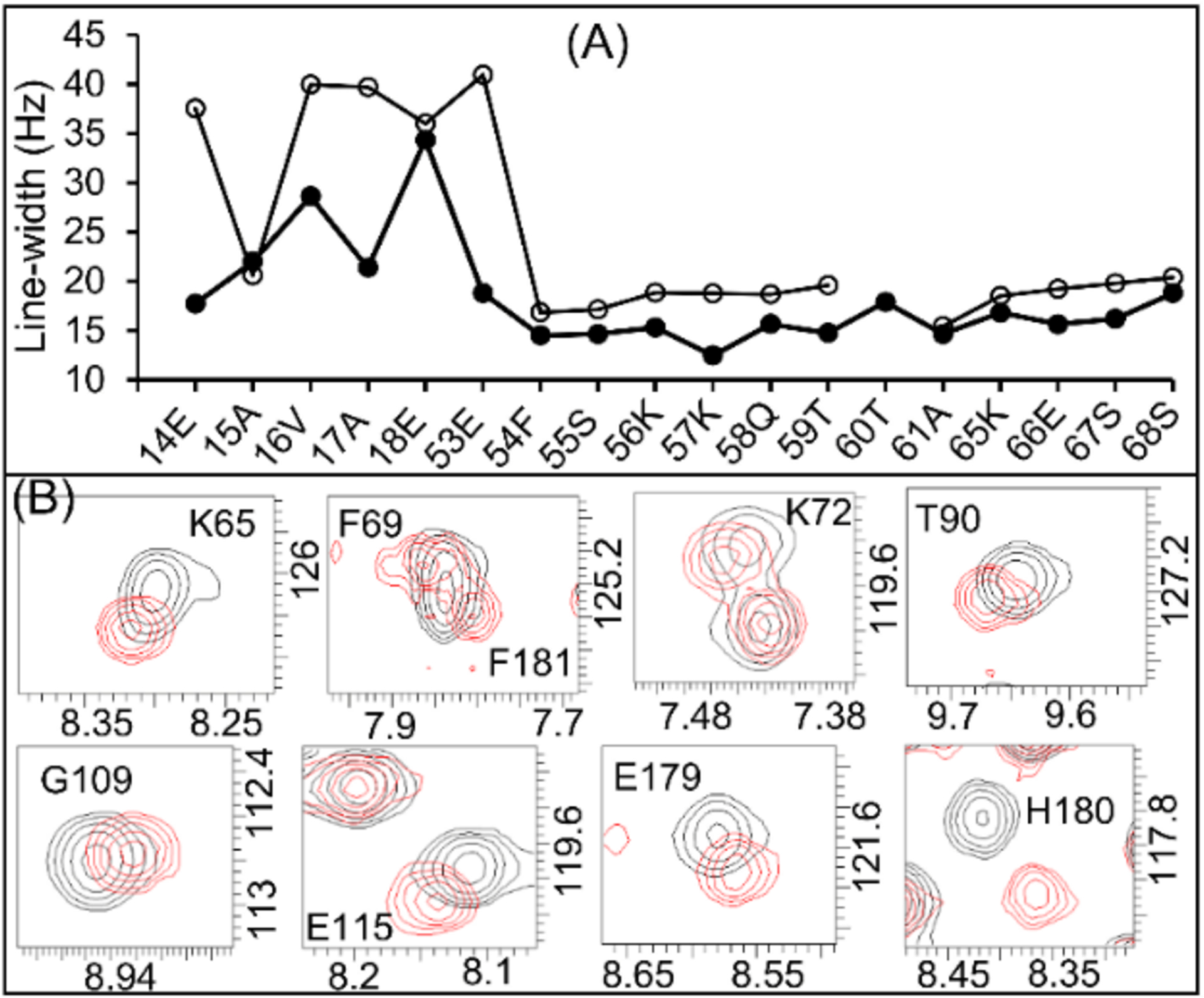
Figure 4.

(A) 1H (open circles) and 15N (closed circles) line-widths of the amino acid residues near to the bilayered lipid-membrane (residues 14–18 and 53) compared to those in the soluble domain (residues 53–68) of the full-length FBD. (B) Expansion of 2D [15N-1H]-HSQC spectra of the full-length (black colored contours) and the truncated (red colored contours) FBD highlighting the perturbed resonances.
