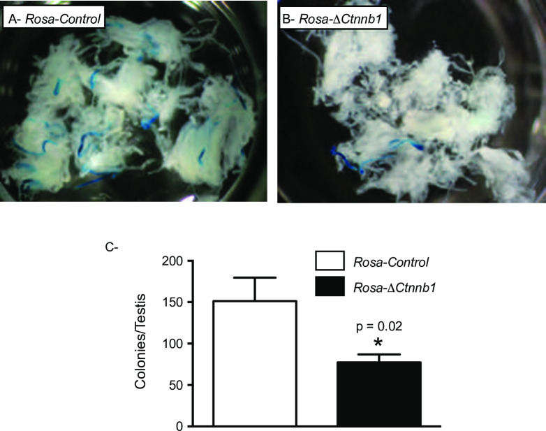
Fig 3. ΔCtnnb1 mice lose spermatogonial stem cell activity.
A, B) Photographs of decapsulated, LacZ-stained recipient testes 8 weeks after transplantation of donor cells from 8-month-old (A) Rosa-control and (B) Rosa-ΔCtnnb1 mice. Blue tubule segments represent colonies of donor-derived spermatogenesis. C) Total numbers of functional spermatogonial stem cells present in a donor testis, calculated by multiplying colony numbers (colonies per million transplanted cells) by the total number of germ cells harvested from a donor testis (n = 4 Rosa-control and n = 5 Rosa-ΔCtnnb1 donor animals whose cells were transplanted in 13 recipient testes/genotype). Data are expressed as means (columns) ± SEM (error bars). * Significant difference from control (P = 0.02).

