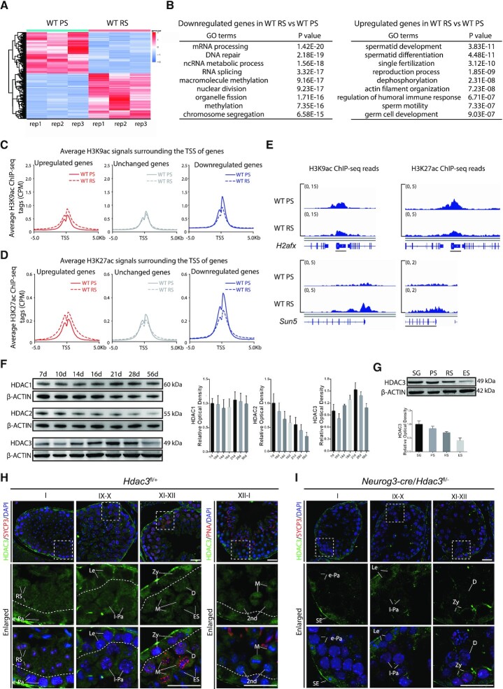
Figure 1.
Transcriptomic changes during the meiotic spermatocytes-to-postmeiotic RS transition are correlated with the genome-wide histone acetylation levels. (A) A heatmap depicting gene expression pattern at round spermatid stage versus pachytene spermatocyte stage in wild-type. Fold change >1.5 up (red) or down (green) with P < 0.01. PS, pachytene spermatocytes; RS, round spermatids. (B) Top nine enriched biological processes for the genes expressed at higher levels or lower levels in RS compared to PS in wild-type. (C) Average H3K9ac profiles at upregulated (red), unchanged (gray), and downregulated (blue) genes in RS versus PS in adult wild-type. Average H3K9ac signals, represented by counts per million, from –5 to + 5 kb surrounding the TSS of genes were shown. (D) Average H3K27ac profiles at upregulated (red), unchanged (gray), and downregulated (blue) genes in RS versus PS in adult wild-type. Average H3K27ac signals, represented by counts per million, from –5 to + 5 kb surrounding the TSS of genes were shown. (E) Genome browser tracks depicting reads accumulation of H3K9ac and H3K27ac on representative meiotic gene H2afx and postmeiotic haploid gene Sun5. (F) Western blot analysis of HDAC1, HDAC2, and HDAC3 during spermatogenesis. β-Actin serves as a loading control. The image is a representation of two independent experiments with similar results. The corresponding optical density readings for biological duplicates are shown. (G) The protein amount of HDAC3 in different fractions of spermatogenic cells. SG: spermatogonia; ES: elongating spermatids. PS, RS, and ES were from mice at age 7–8 weeks (n = 8), and spermatogonia (SG) were isolated from mice at postnatal day 6–8 using the STA-PUT method (n = 20). Quantification of HDAC3 protein with the corresponding optical density readings are shown. The representative image of biological duplicates is shown. (H) Immunofluorescent staining of HDAC3 (green), SYCP3 (lateral elements of the synaptonemal complex, red), and Hoechst (DNA, blue) on frozen sections from adult wild-type. Areas within the rectangles were enlarged in the following panel, and different stages of seminiferous tubules were shown. Abbreviations: SE: somatic Sertoli cells; SG: spermatogonia; RS: round spermatids. Spermatocytes were categorized into the following groups: Le: leptotene; Zy: zygotene; e-Pa: early pachytene; m-Pa: mid pachytene; l-Pa: late pachytene; D: diplotene; M: Metaphase; 2nd: secondary spermatocytes. Scale bars, 25 μm. (I) Immunofluorescent staining of HDAC3 (green), SYCP3 (red) and Hoechst (DNA, blue) on frozen sections from adult Neurog3-cre/Hdac3fl/– testes. Scale bars, 25 μm.

