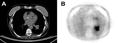
Figure 1 .

(A) Chest CT with contrast enhancement displaying a solid lung mass of 38 mm characterized by polycyclic margins and dysmorphic calcification. (B) PET/CT with fluorodeoxyglucose revealing high glucose metabolism of the lung mass in the perihilar region of the left inferior lung lobe, near the left inferior lobar bronchus.
