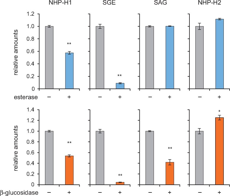
Figure 4.
Effects of esterase and β-glucosidase treatments on NHP-H1, NHP-H2, SGE, and SAG. An extract of Arabidopsis leaves inoculated with Psm that accumulated NHP- and SA-conjugates was buffered to pH 6.0 with 0.1 mM sodium phosphate, and aliquots thereof incubated for 15 h with 10 U mL−1 esterase (+), 10 U mL−1 β-glucosidase (+), or with buffer only (−). Samples were analyzed by GC–MS (Figure 2), and amounts of analytes were related to ribitol as internal standard. All four hexose conjugates were stably detectable by GC–MS when incubated overnight at pH 6.0. Values are expressed relative to the means of the buffer only condition (n = 3). Asterisks indicate significant differences between buffer only (−) and enzyme-treatments (**P < 0.001 and *P < 0.01 (two-tailed Student’s t test).

