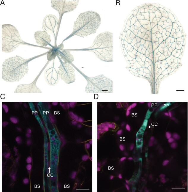
Figure 6.

Reporter gene analysis of pbZIP9:GFP-GUS plants. (A and B) GUS-stained transgenic plants expressing the transcriptional pbZIP9:GFP-GUS reporter construct show GUS activity in the leaf vasculature. Four-week-old plants grown in LD conditions were used for GUS histochemistry. A magnified image of the seventh leaf is shown in b. Scale bars: 1 mm (A) and 0.5 mm (B). (C and D) Confocal microscopy images of two independent pbZIP9:GFP-GUS reporter lines showing GFP fluorescence specific for PP. Magenta, chlorophyll autofluorescence. Yellow, FM4-64FX, Cyan, GFP fluorescence. BS, bundle sheath; CC, companion cell; PP, phloem parenchyma cells are marked. Scale bar: 10 µm.
