Figure 1.
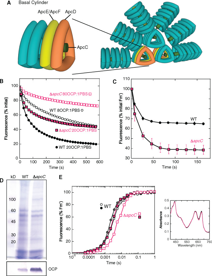
Effect of the lack of ApcC in Synechocystis cells. (A) The Synechocystis PBS. Six PC rods radiate from the core formed by three APC cylinders. A basal cylinder is shown where the position of the trimers containing ApcD or ApcE/ApcF is indicated. The position of ApcC is also given. (B) Purified PBSs (0.012 µM) from WT (black) and ΔapcC mutant (fuchsia) were incubated with two different concentrations of pre-photoactivated OCP giving a final ratio of 20 (closed symbols) or 8 (open symbols) OCP per PBS. The decrease of fluorescence induced in 0.5 M phosphate buffer at 23°C under strong blue light (900 μmol m−2 s−1) is shown (three biologically independent experiments). The error bars are not shown for clarity. (C) Decrease of maximal fluorescence induced by strong blue light in cells. WT (black) and ΔapcC (fuchsia) Synechocystis cells were adapted to low blue light (80 µmol photons s−1 m−2) and then illuminated with strong blue light (1,200 µmol photons s−1 m−2) causing a decrease in fluorescence (Fm′). The ratio PC to chlorophyll was similar in WT and ΔapcC cells. Three biologically independent experiments were performed. The error bars represent sd. The complete PAM traces are shown in Supplemental Figure S1. (D) Coomassie Brilliant Blue and immunoblot detection of OCP in membrane-PBS fractions purified from WT and ΔapcC mutant. For each lane, 2 µg of chlorophyll was loaded. The detection of OCP in other PBS mutants is shown in Supplemental Figure S2. (E) Induction of fluorescence in dark-adapted (closed symbols) and in high-light exposed (open symbols) WT (black squares) and ΔapcC mutant (fuchsia circles) cells in the presence of DCMU (10 µM) and DBMIB (10 µM). A representative experiment is shown. The measurements were repeated with three independent biological replicates. Inset: Absorbance spectra of WT (black) and mutant (fuchsia) cells used in this experiment. The ratio PC to chlorophyll was similar in WT and ΔapcC cells.

