
FIGURE 4.
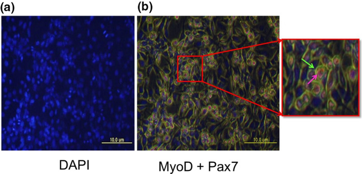
Expression of MyoD and Pax7 in C2C12 mouse myoblasts. C2C12 mouse myoblast cells showed co‐expression of MyoD and Pax7. Cells were immuno‐stained with primary antibodies against MyoD and Pax7. DAPI nuclear staining in represented in (a), and co‐expression of MyoD (green) and Pax‐7 (pink) are shown in (b). Scale bar: 10 μm