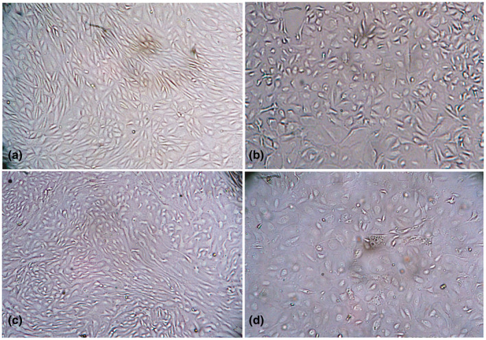
FIGURE 2.

Morphology of PPRV infected primary goat kidney cells at 36 and 48 hr post infection. Uninfected normal (a) and infected goat kidney cells at 36 hr post infection showing rounding of cells (b), uninfected normal (c) and infected goat kidney cells at 48 hr post infection showing vacuolation and syncytia formation (d). Objectives: 20× (a–d)
