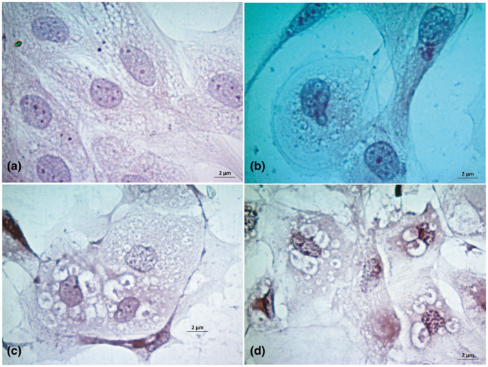
FIGURE 4.

Study of cytopathic effects in goat kidney cells upon H & E stain. Uninfected control goat kidney cells (a), nuclear budding with syncytia formation at 48 hr post infection (b), vacuolation, nuclear budding and syncytia formation at 72 hr post infection (c) and vacuole, syncytia and nuclear budding at 96 hr post infection in goat kidney cells (d) are shown. Bar (=2 µm) indicates the magnification
