
Fig. 3.
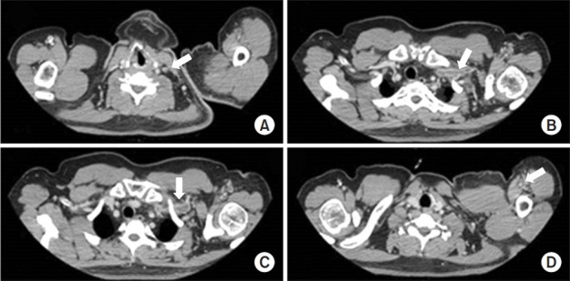
Computed tomography venography showed deep vein thrombosis in the internal jugular vein (A), brachiocephalic vein (B), axillary vein (C), and brachial vein (D) in the left extremity of the patient.
Official websites use .gov
A
.gov website belongs to an official
government organization in the United States.
Secure .gov websites use HTTPS
A lock (
) or https:// means you've safely
connected to the .gov website. Share sensitive
information only on official, secure websites.
Computed tomography venography showed deep vein thrombosis in the internal jugular vein (A), brachiocephalic vein (B), axillary vein (C), and brachial vein (D) in the left extremity of the patient.