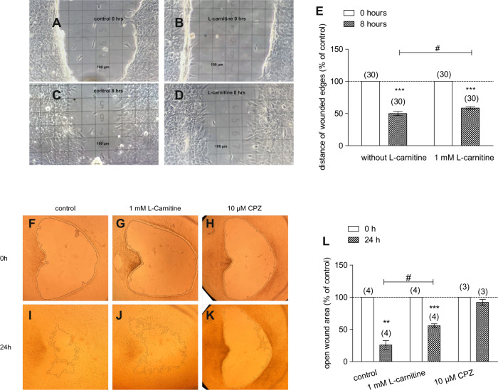
Fig. 6. HCK migration with and without L-carnitine.
When HCK reached confluence, a scratch was created with a pipette tip, and floating cells were removed by washing with PBS. The wounded edges and open wound areas were then observed from 0 to 8 and 24 h of treatment with and without the treatment of 1 mmol/l L-carnitine. Images of cell migration were acquired and microscopic fields of view of representative experiments are shown. A Light microscopic image of HCK at 0 h (control) (square = 100 µm). B HCK in the presence of 1 mmol/l L-carnitine. C HCK (control) after 8 h. D HCK (L-carnitine) after 8 h. E Summary of the experiment regarding distance of wounded edges (control set to 100%). The asterisks (*) designate significant decreases of the distances of the wounded edges (% of control) (n = 30; p < 0.005; paired tested) compared to control. The hashtag (#) indicates statistically significant difference of the wounded edges with and without the treatment of L-carnitine (n = 30; p < 0.05; non-paired tested). F–K Same experimental design as shown in A–D, but with focus on open wound area between 0 and 24 h. The open wound areas were marked by the software. Furthermore, CPZ was used as a negative control. L Summary of the experiments regarding open wound areas (control set to 100%). The asterisks (*) designate significant decreases of the open wound areas (% of control) (n = 3–4; p < 0.01–0.005; paired tested) compared to control. In contrast, the open wound areas did not decrease in the presence of CPZ. The hashtag (#) indicates statistically significant differences in open wound areas (% of control) between control and L-carnitine (t = 24 h; n = 4; p < 0.05, unpaired tested).

