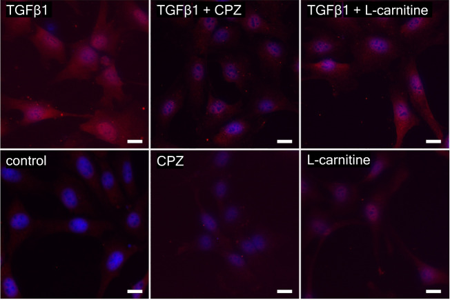
Fig. 7. Myofibroblast activation in HCK after stimulations for 24 h with 5 ng/ml human recombinant transforming growth factor beta 1 (TGFβ1), 10 µmol/l capsazepine (CPZ), 1 mmol/l L-carnitine, 5 ng/ml TGFβ1 plus 10 µmol/l CPZ, 5 ng/ml TGFβ1 plus 1 mmol/l L-carnitine and, as a control, with Dulbecco’s Modified Eagle Medium (DMEM).
Myofibroblasts become apparent as cells that stain positive for αSMA in the cytoplasm. Nuclear staining of HCK with DAPI (blue) and anti αSMA IF antibody red staining detecting myofibroblasts in HCK. Scale bar = 20 µm.

