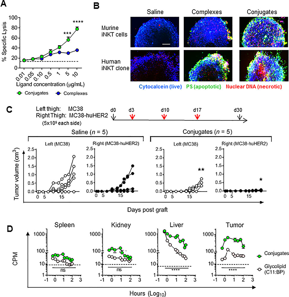
Figure 4; Specific cytotoxicity and tissue deposition of HER2-targeted conjugates.
(a) MC38-huHER2 cells labeled with 51Cr labeled were incubated with anti-huHER2 conjugates or complexes. Target specific killing was determined following incubation for 12 hr with human iNKT cell clone HDD3 at effector:target ratio of 1:1. (b) Spheroids of MC38-huHER2 cells were pulsed with saline or anti-huHER2 complexes or conjugates containing either murine (top) or human (bottom) CD1d. Following incubation for 12 hr with mouse spleen iNKT cells (top) or human iNKT cells (clone HDD3, bottom), spheroids were stained for markers of viability, apoptosis and necrosis. Scale bar 1 mm. (c) Mice were grafted on day 0 with 5 × 10⁵ MC38-huHER2 cells s.c. in the right thigh and 5 × 10⁵ huHER2 negative MC38 cells in the left thigh. Injections of saline or anti-huHER2 conjugates were given i.v. on days 3, 10 and 17, and growth of tumors on left and right thighs was followed to day 30. (d) Mice grafted with 5 × 10⁵ MC38-huHER2 tumor cells 14 days earlier received a single i.v. injection of 1 nanomole of anti-HER2 conjugates loaded with 14C-labelled C11:BP glycolipid or 1 nanomole of 14C-labeled free C11:BP glycolipid. Individual mice were sacrificed at times shown, and 14C retention in organ or tumor tissues was determined by liquid scintillation counting. Dashed lines indicate background CPM for mice injected with saline only. *P < 0.05, **P < 0.01 ***P < 0.001, ****P < 0.0001, 2-way ANOVA with Sidak multiple comparison test.

