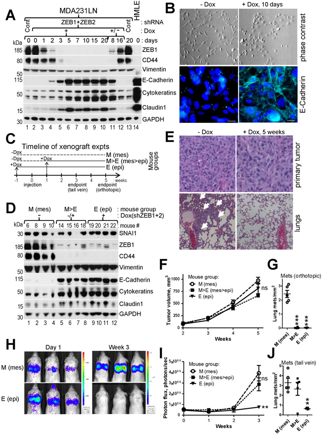
Fig.7. Suppression of ZEB1+2 and EMT inhibits lung metastasis at multiple stages.
A, WB analysis of the indicated EMT markers in MDA231LN cells during time-course induction of Dox-regulated shRNAs against ZEB1+2. Cells were lysed at the indicate time points post Dox addition (lanes 1-10,13). Dox was washed out on day 20 and the cells were lysed 8 and 16 days post Dox withdrawal (lanes 11,12). B, Phase contrast images (top panel) and immunofluorescence for E-Cadherin pseudocolored cyan (bottom panel) of cells shown in (A). C, Timeline for mouse groups M, M>E and E used in xenograft experiments described in (D-J). D-J, MDA231LN/shZEB1+2 cells described in (A&B) were injected into mammary fad pad (D-G), or into tail vein (H-J) of NSG mice. D, WB analysis of the indicated EMT markers in primary tumors of four mice from each of the three groups at endpoint of the orthotopic injection experiment. E, H&E staining of primary tumors (top panel) and lungs (bottom panel) from (D). White arrows point to macrometastases. F-J, Dynamics of tumor growth (F and I) and the number of lung metastases at endpoints (G and J) for orthotopic and intravenous injection experiments, respectively (n=5 mice per group). H, Representative bioluminescent images for the tail vein injection. The data are mean ± SEM. ANOVA with Dunnett’s post-test, * - p<0.05, ** - p<0.01, *** - p<0.001, ns – non-significant.

