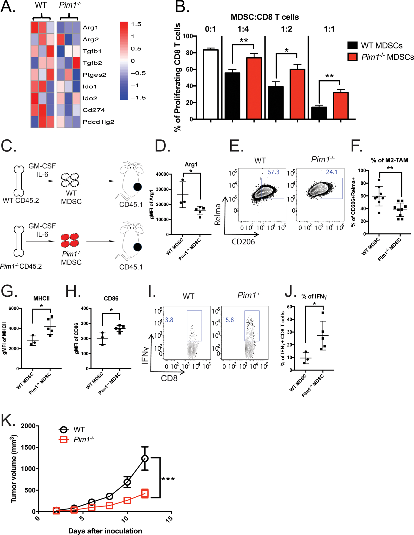
Figure 3. PIM1 is required for the immunosuppressive phenotype of MDSCs.

(A) Heatmap showing expression of genes associated with immunosuppressive functions in BM-MDSCs generated from WT (n=3) and Pim1−/− (n=3) mice. (B) WT (n=3) and Pim1−/− (n=3) MDSC suppressive activities were evaluated by the ability to inhibit CD8+ T-cell proliferation. Bar graphs were plotted from three independent experiments. (C-K) WT (n=3) and Pim1−/− (n=5) BM-MDSCs were purified and transferred into CD45.1+ congenic recipient mice bearing an MC38 tumor. (C) Experimental design. (D) Levels of Arg1 protein expression in intratumoral myeloid cells derived from WT and Pim1−/− donor cells. (E) Representative plot showing the frequencies of M2 macrophages evaluated by CD206 and Relmα expression from WT and Pim1−/− donor cells. (F) Quantification of E. (G-H) Protein expression levels of MHCII and CD86. (I-J) Intratumoral CD8 T cells from WT and Pim1−/− MDSC recipients were stimulated and evaluated for IFNγ production by flow cytometry as shown in representative (I) and quantitative (J) plots. (K) Tumor growth was measured using calipers for two weeks and plotted. Data are expressed as mean ± SEM from two experiments with 3 mice per group and significance was determined by t-test. * p < 0.05, ** p < 0.01, *** p < 0.001.
